
Thu, Aug 27, 2026

Volume 20, Issue 2 (Mar-Apr 2026)
mljgoums 2026, 20(2): 36-41 |
Back to browse issues page


Download citation:
BibTeX | RIS | EndNote | Medlars | ProCite | Reference Manager | RefWorks
Send citation to:


BibTeX | RIS | EndNote | Medlars | ProCite | Reference Manager | RefWorks
Send citation to:
Behfar B, Haddadi F, Sharifmoghadam M R, Kamaladini H, Bahreini M, Niknejad A. Characterization and optimization of keratinase production by bacillus pumilus and bacillus tequilensis isolated from poultry waste. mljgoums 2026; 20 (2) :36-41
URL: http://mlj.goums.ac.ir/article-1-1787-en.html
URL: http://mlj.goums.ac.ir/article-1-1787-en.html
Bahareh Behfar1 

 , Fatemeh Haddadi2 , Mohammad Reza Sharifmoghadam3 , Hossein Kamaladini1 , Masoumeh Bahreini3 , Azadeh Niknejad4
, Fatemeh Haddadi2 , Mohammad Reza Sharifmoghadam3 , Hossein Kamaladini1 , Masoumeh Bahreini3 , Azadeh Niknejad4
1- Department of Biology, Faculty of Sciences, University of Zabol, Zabol, Iran
2- Department of Biology, Faculty of Sciences, University of Zabol, Zabol, Iran ,haddadifatemeh@yahoo.com
3- Department of Biology, Faculty of Sciences, Ferdowsi University of Mashhad, Mashhad, Iran
4- Department of Cellular and Molecular, Faculty of Biological Sciences, Kharazmi University, Tehran, Iran
2- Department of Biology, Faculty of Sciences, University of Zabol, Zabol, Iran ,
3- Department of Biology, Faculty of Sciences, Ferdowsi University of Mashhad, Mashhad, Iran
4- Department of Cellular and Molecular, Faculty of Biological Sciences, Kharazmi University, Tehran, Iran
Full-Text [PDF 561 kb]
(1563 Downloads)
| Abstract (HTML) (6403 Views)
Full-Text: (639 Views)
Introduction
Keratin is an insoluble and highly stable protein because of the high content of disulfide bonds in its structure, which tightly connects the peptide chains and makes it resistant to enzymatic digestion (1). Keratin is a highly cross-linked, fibrous protein that forms the structural component of hair, feathers, nails, horns, and other biological materials (2). Keratin is structurally divided into two groups: α-keratin and β-keratin. α-Keratin has a right-handed helix structure and is mainly found in hair and wool. In contrast, chicken feathers are mainly composed of β-helix and are classified as β-keratin (3). Keratin is also classified as hard keratin (Feathers) and soft keratin (Epidermis), depending on its sulfur content (4). In nature, keratin waste is hydrolyzed by a wide range of microorganisms as a source of energy, thereby reducing pollution (5).
Millions of tons of keratinous waste, including chicken feathers, are produced annually in various industries worldwide. Because of their impurities, disposal of these wastes has an adverse effect on soil fertility. The release of contaminants such as arsenic into poultry litter is also a major concern (6). However, these wastes are rich sources of keratin fiber (82%) (7), elements, vitamins, and growth factors (8), proteins, peptides, and amino acids (9), and they are mostly either discarded or burned (10), causing the growth of pathogenic microorganisms and air pollution (11). Keratinase is a protease that mostly attacks disulfide bonds in the keratin structure (12). Microorganisms, such as yeast and bacteria, are able to produce keratinolytic enzymes that can hydrolyze the highly stable protein keratin into simpler forms (13). Therefore, keratinase is a potential enzyme that can be used in animal feed, biomedicine, detergents, cosmetics, the textile and leather industries, biofuels, and bioremediation (14). Biodegradation of chicken waste by the enzyme keratinase is an environmentally friendly and cost-effective ecological process that can play an important role in biotechnological applications (15).
The recognition of keratinolytic microorganisms was first reported by Noval and Nickerson (16). The resistance of keratin to some proteases, such as papain, pepsin, and trypsin, was reported in 1986, and keratinases are the only group of proteases capable of complete hydrolysis of these insoluble and complex proteins (17). Keratinolytic bacterial isolates have been mainly documented among strains of the Gram-positive genera Bacillus (18) and Streptomyces (19), while some studies suggest keratinolytic properties in some Gram-negative bacteria (20). The degradation process of natural keratin is enhanced by the addition of sulfide reductase. The initial action of keratinase on the keratin substrate does not change significantly for two to four hours but is sufficient for further hydrolysis by trypsin and pepsin proteases (5). The most important application of keratinases is the biological disposal of waste by decomposing keratin and other resistant materials, such as gelatin, elastin, fibrin, and collagen (5).
In addition to removing contamination, several applications have been investigated, including drug manufacturing, weeding, textiles, production of organic fertilizers for soil remediation (15), leather soaking, X-ray film recycling (15), topical treatment of nail diseases, and hard keratin depletion (4), as well as the production of various materials, such as detergents for removal of egg stains and blood (21), anti-dandruff shampoos (22), green energy biofuels, and insecticides and nematode suppressors (23).
The primary focus of this investigation was to identify and explore the potential of microorganisms capable of degrading keratin, a resilient structural protein that is found in abundance in poultry waste. Toward this end, two keratinolytic isolates, designated FUM120 and FUM122, were obtained from poultry waste samples collected around the city of Mashhad. These isolates were subjected to comprehensive quantitative and qualitative examination in the Microbiology Department of the Ferdowsi University of Mashhad. The characterization process involved a combination of biochemical and molecular techniques, including identification of the 16SrRNA and keratinase gene sequences for both isolates, which were subsequently deposited in the NCBI database.
Furthermore, the study sought to thoroughly characterize the isolated microbes through a combination of morphological, biochemical, and molecular techniques. Additionally, the research aimed to optimize the culture conditions to facilitate the production of keratinase, the key enzyme involved in the breakdown of keratin. Lastly, the study evaluated the keratinolytic activity exhibited by the isolated microorganisms, with the goal of identifying promising candidates for potential applications in waste management or other industrial processes.
Methods
All chemicals and culture media were obtained from Sigma-Aldrich Co., Germany.
In this study, keratinolytic bacteria were isolated from poultry waste around Mashhad (Iran) in the Microbiology Department of the Ferdowsi University of Mashhad. Initial screening was performed using a specialized solid feather meal agar (FMA) culture medium. The bacterial isolates were cultured on nutrient agar plates at 37°C overnight and then maintained at 4°C.
Motility was tested in SIM medium. Morphology was identified using the Gram-staining procedure.
Urease-producing capacity was investigated using urea broth medium containing phenol red. Hydrolysis of H2O2 by catalase was tested near the isolate colony using a 3% aqueous solution of H2O2. The growth of isolates was examined in Simmons’ citrate agar containing sodium citrate as the only carbon source (24). Kovac’s reagent was used to determine the presence of indole (25). H2S production was investigated by the formation of an insoluble black precipitate in the SIM medium. Gelatinase activity was determined in basal medium agar supplemented with gelatin after 24 hours of incubation at 37°C. Voges-Proskauer (26) and methyl red tests were performed in MR-VP broth to measure the fermentation of butanediol and glucose, respectively. Nitrate reduction was investigated in a medium supplemented with KNO3 (27). The ability to produce amylase was assayed in a medium containing 1% starch using Lugol’s iodine solution. Growth of the isolate was assessed in litmus milk for 24 - 48 hours at 37°C (28). The isolate was cultured on nutrient agar supplemented with 3%, 6%, and 10% NaCl for 24 - 48 hours at 37°C. Antibiotic sensitivity to amikacin, ampicillin, gentamicin, and tetracycline was evaluated on Mueller-Hinton agar. Fermentative-oxidative metabolism was also tested.
The genomic DNA of FUM120 and FUM122 was extracted using a kit from ZandBiotech Co., Iran. The 16SrRNA sequences were amplified by universal primers (Synthesized by Sinacloon, Iran), forward 5´-AGAGTTTGATCMTGGCTCAG-3´ and reverse 5´-GGTTACCTTGTTACGACTT-3´, and Taq DNA Polymerase 2x Master Mix Red (Ampliqon). The thermal cycler was set up as follows: 95°C for 10 minutes as the initial denaturation step, 35 cycles of 95°C for 30 s, 56°C for 60 s, 72°C for 60 s, and finally 72°C for 10 minutes as the final extension step. The amplified fragment was purified from agarose gel using a kit from ZandBiotech Co., Iran, and was sequenced at the School of Health, National Influenza Center, University of Tehran. The sequencing results were analyzed using the BLAST tool of the NCBI GenBank database. The phylogenetic relationship of the FUM120 and FUM122 isolates was determined following the neighbor-joining tree method with bootstrap values for 500 replicates using molecular evolutionary genetics analysis (MEGA) X (Version 10.0) software (29).
The industrial feathers were treated with detergents for several hours to remove lipids and preservatives. The washed feathers were dried at 37°C for 24 hours and then pulverized using a meal mixer.
A single colony from the keratinolytic isolates was cultured in Tryptic Soy Broth (TSB) and incubated at 37°C and 200 rpm for 18 hours. Then, 2% (v/v) of prepared 0.5 McFarland from the overnight culture was transferred into feather meal agar (FMA) containing 1% feather powder as the only carbon, nitrogen, and sulfur source, 0.05% NaCl, 0.04% KH2PO4, 0.03% K2HPO4, 0.01% MgSO4, and NH4Cl in 100 mL Erlenmeyer flasks with a volume of 50 mL. After five days of incubation at 37°C and 150 rpm, 2 mL of culture was centrifuged at 10000 rpm for 5 minutes, and the supernatant was assayed for keratinase activity.
Fifteen microliters from the overnight culture of the keratinolytic isolate with a standard concentration of 0.5 McFarland were cultured on skim milk agar and transferred to the incubator at 37°C for 24 hours. A clear zone around the isolate growth site was indicative of proteolytic activity. To investigate the keratinolytic properties, the isolate was cultured on FMA agar containing 1% feather powder as the only source of carbon and nitrogen. The growth of the isolate and formation of a clear zone around it indicated keratinase activity.
Azokeratin was prepared as described by Tomarelli et al. (30), then freeze-dried and stored at 4°C. For the keratinase assay, 10 mg azokeratin was dissolved in 0.9 mL of 50 mM Tris-HCl (pH 8.0); 0.1 mL enzyme from the centrifuged supernatant was mixed with the azokeratin, and the mixture was incubated for 60 minutes at 37°C in a shaker incubator. The reaction was terminated by placing it on ice for half an hour, and then it was filtered. Finally, the absorption of the filtered solution was measured at 450 nm. The bacteria-free culture medium was used as a negative control. One unit of enzyme activity was defined as a 0.1 increase in absorbance at 450 nm per mL after 60 minutes of incubation.
Similar to the enzymatic assay with azokeratin, the mixture assay was prepared with 50 mg feather powder. After incubation at 37°C for 60 minutes, 0.2 mL 10% TCA was added to the mixture, and it was maintained at -20°C for 10 minutes. After centrifugation at 14000 rpm for 15 minutes, the absorbance was measured at 280 nm.
Results
The isolated keratinolytic strains were studied based on cell morphology, spore production, Gram staining, several biochemical tests, and 16SrRNA sequencing. Tables 1 and 2 summarize the morphology and biochemical characteristics of the isolates. The FUM120 isolate was Gram-positive, sporulating, and shaped as a short rod. The growth of this bacterium on nutrient agar formed small colonies. Similar to the first isolate, the FUM122 isolate was a Gram-positive and rod-shaped bacterium that produced spores. This isolate formed large mucoid colonies on nutrient agar (Figure 1A-C). Antibiotic sensitivity of the isolates was investigated against amikacin, ampicillin, gentamicin, and tetracycline. Despite the sensitivity of FUM122 to all antibiotics, FUM120 was resistant to ampicillin, indicating possible beta-lactamase production (Table 2).
Table 1. Results of biochemical characterization of FUM120 and FUM122 isolates
.PNG)
The nucleotide sequence of the keratinase-producing isolates was amplified to a length of 1500 bp (Figure 2). Analysis of the 16SrRNA nucleotide sequences of the isolates with the Basic Local Alignment Search Tool (BLAST) indicated that FUM120 was approximately 100% similar to Bacillus pumilus, while the FUM122 isolate was mainly similar to Bacillus tequilensis. These results were confirmed by the biochemical test results. The 16SrRNA gene sequences of Bacillus pumilus FUM120 and Bacillus tequilensis FUM122 were submitted to GenBank with accession numbers MT062864.1 and MT062878.1, respectively. The phylogenetic relationships of the isolates were constructed based on BLAST alignment to GenBank (Figure 3). Bootstrap analysis showed that the FUM120 isolate was in the Bacillus pumilus branch, while FUM122 was in the Bacillus tequilensis branch.
Table 2. Results of antibiotic sensitivity tests on FUM120 and FUM122 isolates
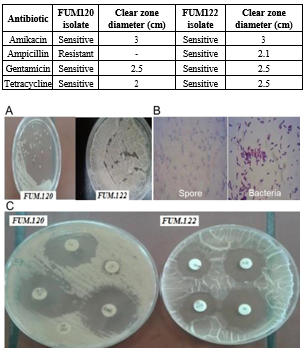
Figure 1. The bacterial identification: (A) Colony morphology in the nutrient agar, (B) Bacterial morphology under microscope, (C) Results of antibiotic sensitivity tests on FUM120 and FUM122 isolates
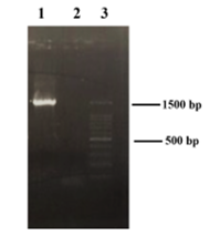
Figure 2. The nucleotide sequence of keratinase-producing isolates was amplified to a length of 1500 bp (1), negative control (2), ladder (3)
Protease production was investigated by bacterial growth on a skim milk plate, and the keratinolytic properties of the proteases were confirmed using FMB containing feather powder as the only carbon, nitrogen, and sulfur source (Figure 4 A and C) for 24 hours at 37°C. Clear zone formation around the isolates’ colonies in skim milk indicated protease secretion, which was 18.67 and x mm for Bacillus pumilus FUM120 and Bacillus tequilensis FUM122, respectively. Through hydrolysis of feather keratin, keratinase enabled the bacterium to grow in the FMB. The activity of extracellular keratinase of Bacillus pumilus FUM120 was 32.29 U mL-1 during five days of cultivation using azokeratin as the substrate (Figure 4B). The keratinase activity of Bacillus tequilensis FUM122 was measured as x U mL-1 under the same conditions (Figure 4, Table 3).

Figure 3. Neighbor-joining phylogenetic relationships of Bacillus pumilus FUM120 (A) and Bacillus tequilensis FUM122 (B) by bootstrap value for 500 replicates based on 16SrRNA gene sequencing to GenBank

Figure 4. Investigation of enzyme activity with three methods: (A) The formation of the clear zone in skim milk medium, (B) Azokeratin reagent, (C) Hydrolysis of the feather in FMB
Table 3. Quantitative measurement of keratinase activity

Discussion
The isolation and characterization of keratinase-producing bacteria have received increasing attention in recent years, with a focus on efficient and sustainable methods for utilizing keratin-rich wastes. In the present study, the isolate was identified as a Bacillus sp. based on morphological, biochemical, and 16SrRNA gene sequence analysis.
The optimization of culture conditions, such as pH, temperature, and incubation time, is crucial for enhancing keratinase production. In this study, the maximum keratinase activity was observed at pH 7.0 and 37°C, consistent with reported optimal conditions for Bacillus species. For instance, Bacillus licheniformis ALW1 showed optimal keratinase activity at pH 8.0 and 65°C (31), while Bacillus aerius NSMk2 exhibited the highest keratinase production at pH 7.5 and 35°C (32). These differences reflect the strain-specific nature of keratinase production and the need for optimization for each isolate.
Keratinaceous byproducts, such as feathers, hairs, nails, and horns, are significant wastes that require management. Feather wastes can be decomposed by microorganisms with enzymes that hydrolyze keratin, saving energy and preventing amino acid degradation (1). This approach is preferable to traditional chemical treatment methods, which are energy-intensive and may cause amino acid loss.
Keratinolytic enzymes are produced by many microorganisms, including bacteria, actinomycetes, and fungi, which are often isolated from poultry waste environments (32-34). Among bacteria, keratinase production is mainly associated with Gram-positive species, particularly the Bacillus genus (35-38), although some Gram-negative bacteria, such as Vibrio, Xanthomonas, and Chryseobacterium, also produce keratinases (20).
Various Bacillus species have been extensively studied for keratinase production, and optimization strategies have been employed to enhance yields. For instance, Bacillus licheniformis, B. aerius, and B. zhangzhouensis produce keratinases with optimal activities under diverse pH and temperature conditions. The optimization of culture media, including suitable carbon and nitrogen sources, has been crucial for improving keratinase production.
Bacillus sp. CL33A keratinase was produced in a medium with feather meal as a low-cost substrate. Keratinolytic activity was optimized at 48 - 62°C and a pH of 7.2 - 9.2 (39). Bacillus licheniformis strain NBRC 14206 produces a keratinase that is most stable at alkaline pH and 40°C. The keratinolytic activity of purified enzymes was slightly stimulated by Ca2+, Zn2+, and Fe2+, while EDTA had a significant stimulatory effect on activity (40). Bacillus aerius NSMk2 showed the highest keratinase production in the minimal salt medium supplemented with fructose and beef extract and improved degradation of chicken feathers. The maximum keratinase activity was obtained at 1.375%, a pH of 7.5, and a temperature of 35°C in two days, sevenfold that in minimal salt medium (41). The keratinase obtained from Bacillus licheniformis ALW1 in optimized medium increased biosynthesis to 72.2 U/mL (2.9-fold) at a pH of 8.0 and a temperature of 65°C with 0.7% soluble keratin. This enzyme showed stability at 50 - 60°C and alkaline pH for 90 minutes (31). Bacillus licheniformis BBE11-1 and Stenotrophomonas maltophilia BBE11-1 were co-cultured in a medium with 50 g/L chicken feather waste, and the degradation rates improved to 55% in 96 hours compared to single cultivation. Optimization of the co-culture conditions increased the degradation rate to 81.8% (42). Bacillus sp. Nnolim-K1 keratinase production was optimized in a medium with 0.8% (w/v) xylose, 1% (w/v) feather, and 3% (v/v) inoculum size at a pH of 5 and 25°C. Maximum activity, 1943.43 ± 0.0 U/mL, was obtained after 120 hours under optimum conditions, i.e., pH of 8.0 and 60°C. Keratinase inhibition in the presence of EDTA and 1,10-phenanthroline indicated a metallo-keratinase nature (43). Optimization of Bacillus zhangzhouensis keratinase production doubled enzyme yield. The keratinase w::as char::acterized as a serine protease with a molecular weight of 42 kDa and maximum activity at an alkaline pH and a temperature of 60°C (44). In 2020, many keratinase-producing isolates from Arthrobacter sp. (45) and fungal species, such as Aphanoascus keratinophilus and Chrysosporium tropicum, were introduced, and their production conditions were optimized.
In recent years, Bacillus tequilensis has been reported as a potential species for keratinase production (21-46). In this study, both isolated bacteria showed keratinolytic activity. According to morphological, biochemical, and molecular characterization described in Bergey’s Manual, Bacillus pumilus and Bacillus tequilensis were identified. This study demonstrates the isolation and characterization of a keratinase-producing Bacillus sp. with potential applications in the valorization of keratin-rich wastes and leather processing. The enzyme’s effectiveness in feather degradation and dehairing of goat skin suggests industrial applications. Further optimization and scale-up studies, evaluating enzyme stability and performance under industrial conditions, are necessary to assess commercial viability and support sustainable waste management strategies.
Conclusion
In this study, according to cell morphology, spore formation, Gram staining, biochemical evaluations, and 16SrRNA sequencing, FUM 120 and FUM 122 strains isolated from poultry manure were identified. The strains’ antibiotic sensitivity was reviewed, and BLAST revealed that FUM 120 strain had roughly 100% homology with Bacillus pumilus, while FUM 122 strain had nearly 98% homology with Bacillus tequilensis. Biochemical studies verified these findings. Growth-associated protease synthesis was examined in skim milk, and keratinolytic characteristics were calculated following growth in FMB special media for 24 hours at 37°C using two methods, azokeratin reagent and turbidity calculation.
The detailed characterization and assessment of the keratinolytic properties of FUM 120 and FUM 122 provide insights into potential applications in keratin-rich waste management, textile processing, and other biotechnological domains. Further optimization and scale-up studies are necessary to fully harness their potential.
Acknowledgement
We are very grateful to the Department of Biology, University of Zabol, and the Microbiology Department of the Ferdowsi University of Mashhad for providing laboratory equipment.
Funding Sources
We are very grateful to the University of Zabol for providing grant No. uoz.ac.12/2402.
Ethical Statement
None.
Conflicts of Interest
The authors have no conflict of interest to declare.
Author Contributions
Behfar, B. performed the research, analyzed the data, and provided the initial draft of the manuscript; Haddadi, F. and Sharifmoghadam, M.R. designed the research; Haddadi, F. analyzed the data and wrote and edited the original draft; Haddadi, F., Sharifmoghadam, M.R., Kamaladini, H., and Bahreini, M. provided important supervision of the research. Niknejad, A. analyzed the data and reviewed and edited the manuscript. All authors read and approved the final manuscript.
Data Availability Statement
The datasets used and/or analyzed during the current study are available from the corresponding author upon reasonable request.
Use of Artificial Intelligence
No AI tools were used at any stage of this work.
Keratin is an insoluble and highly stable protein because of the high content of disulfide bonds in its structure, which tightly connects the peptide chains and makes it resistant to enzymatic digestion (1). Keratin is a highly cross-linked, fibrous protein that forms the structural component of hair, feathers, nails, horns, and other biological materials (2). Keratin is structurally divided into two groups: α-keratin and β-keratin. α-Keratin has a right-handed helix structure and is mainly found in hair and wool. In contrast, chicken feathers are mainly composed of β-helix and are classified as β-keratin (3). Keratin is also classified as hard keratin (Feathers) and soft keratin (Epidermis), depending on its sulfur content (4). In nature, keratin waste is hydrolyzed by a wide range of microorganisms as a source of energy, thereby reducing pollution (5).
Millions of tons of keratinous waste, including chicken feathers, are produced annually in various industries worldwide. Because of their impurities, disposal of these wastes has an adverse effect on soil fertility. The release of contaminants such as arsenic into poultry litter is also a major concern (6). However, these wastes are rich sources of keratin fiber (82%) (7), elements, vitamins, and growth factors (8), proteins, peptides, and amino acids (9), and they are mostly either discarded or burned (10), causing the growth of pathogenic microorganisms and air pollution (11). Keratinase is a protease that mostly attacks disulfide bonds in the keratin structure (12). Microorganisms, such as yeast and bacteria, are able to produce keratinolytic enzymes that can hydrolyze the highly stable protein keratin into simpler forms (13). Therefore, keratinase is a potential enzyme that can be used in animal feed, biomedicine, detergents, cosmetics, the textile and leather industries, biofuels, and bioremediation (14). Biodegradation of chicken waste by the enzyme keratinase is an environmentally friendly and cost-effective ecological process that can play an important role in biotechnological applications (15).
The recognition of keratinolytic microorganisms was first reported by Noval and Nickerson (16). The resistance of keratin to some proteases, such as papain, pepsin, and trypsin, was reported in 1986, and keratinases are the only group of proteases capable of complete hydrolysis of these insoluble and complex proteins (17). Keratinolytic bacterial isolates have been mainly documented among strains of the Gram-positive genera Bacillus (18) and Streptomyces (19), while some studies suggest keratinolytic properties in some Gram-negative bacteria (20). The degradation process of natural keratin is enhanced by the addition of sulfide reductase. The initial action of keratinase on the keratin substrate does not change significantly for two to four hours but is sufficient for further hydrolysis by trypsin and pepsin proteases (5). The most important application of keratinases is the biological disposal of waste by decomposing keratin and other resistant materials, such as gelatin, elastin, fibrin, and collagen (5).
In addition to removing contamination, several applications have been investigated, including drug manufacturing, weeding, textiles, production of organic fertilizers for soil remediation (15), leather soaking, X-ray film recycling (15), topical treatment of nail diseases, and hard keratin depletion (4), as well as the production of various materials, such as detergents for removal of egg stains and blood (21), anti-dandruff shampoos (22), green energy biofuels, and insecticides and nematode suppressors (23).
The primary focus of this investigation was to identify and explore the potential of microorganisms capable of degrading keratin, a resilient structural protein that is found in abundance in poultry waste. Toward this end, two keratinolytic isolates, designated FUM120 and FUM122, were obtained from poultry waste samples collected around the city of Mashhad. These isolates were subjected to comprehensive quantitative and qualitative examination in the Microbiology Department of the Ferdowsi University of Mashhad. The characterization process involved a combination of biochemical and molecular techniques, including identification of the 16SrRNA and keratinase gene sequences for both isolates, which were subsequently deposited in the NCBI database.
Furthermore, the study sought to thoroughly characterize the isolated microbes through a combination of morphological, biochemical, and molecular techniques. Additionally, the research aimed to optimize the culture conditions to facilitate the production of keratinase, the key enzyme involved in the breakdown of keratin. Lastly, the study evaluated the keratinolytic activity exhibited by the isolated microorganisms, with the goal of identifying promising candidates for potential applications in waste management or other industrial processes.
Methods
All chemicals and culture media were obtained from Sigma-Aldrich Co., Germany.
In this study, keratinolytic bacteria were isolated from poultry waste around Mashhad (Iran) in the Microbiology Department of the Ferdowsi University of Mashhad. Initial screening was performed using a specialized solid feather meal agar (FMA) culture medium. The bacterial isolates were cultured on nutrient agar plates at 37°C overnight and then maintained at 4°C.
Motility was tested in SIM medium. Morphology was identified using the Gram-staining procedure.
Urease-producing capacity was investigated using urea broth medium containing phenol red. Hydrolysis of H2O2 by catalase was tested near the isolate colony using a 3% aqueous solution of H2O2. The growth of isolates was examined in Simmons’ citrate agar containing sodium citrate as the only carbon source (24). Kovac’s reagent was used to determine the presence of indole (25). H2S production was investigated by the formation of an insoluble black precipitate in the SIM medium. Gelatinase activity was determined in basal medium agar supplemented with gelatin after 24 hours of incubation at 37°C. Voges-Proskauer (26) and methyl red tests were performed in MR-VP broth to measure the fermentation of butanediol and glucose, respectively. Nitrate reduction was investigated in a medium supplemented with KNO3 (27). The ability to produce amylase was assayed in a medium containing 1% starch using Lugol’s iodine solution. Growth of the isolate was assessed in litmus milk for 24 - 48 hours at 37°C (28). The isolate was cultured on nutrient agar supplemented with 3%, 6%, and 10% NaCl for 24 - 48 hours at 37°C. Antibiotic sensitivity to amikacin, ampicillin, gentamicin, and tetracycline was evaluated on Mueller-Hinton agar. Fermentative-oxidative metabolism was also tested.
The genomic DNA of FUM120 and FUM122 was extracted using a kit from ZandBiotech Co., Iran. The 16SrRNA sequences were amplified by universal primers (Synthesized by Sinacloon, Iran), forward 5´-AGAGTTTGATCMTGGCTCAG-3´ and reverse 5´-GGTTACCTTGTTACGACTT-3´, and Taq DNA Polymerase 2x Master Mix Red (Ampliqon). The thermal cycler was set up as follows: 95°C for 10 minutes as the initial denaturation step, 35 cycles of 95°C for 30 s, 56°C for 60 s, 72°C for 60 s, and finally 72°C for 10 minutes as the final extension step. The amplified fragment was purified from agarose gel using a kit from ZandBiotech Co., Iran, and was sequenced at the School of Health, National Influenza Center, University of Tehran. The sequencing results were analyzed using the BLAST tool of the NCBI GenBank database. The phylogenetic relationship of the FUM120 and FUM122 isolates was determined following the neighbor-joining tree method with bootstrap values for 500 replicates using molecular evolutionary genetics analysis (MEGA) X (Version 10.0) software (29).
The industrial feathers were treated with detergents for several hours to remove lipids and preservatives. The washed feathers were dried at 37°C for 24 hours and then pulverized using a meal mixer.
A single colony from the keratinolytic isolates was cultured in Tryptic Soy Broth (TSB) and incubated at 37°C and 200 rpm for 18 hours. Then, 2% (v/v) of prepared 0.5 McFarland from the overnight culture was transferred into feather meal agar (FMA) containing 1% feather powder as the only carbon, nitrogen, and sulfur source, 0.05% NaCl, 0.04% KH2PO4, 0.03% K2HPO4, 0.01% MgSO4, and NH4Cl in 100 mL Erlenmeyer flasks with a volume of 50 mL. After five days of incubation at 37°C and 150 rpm, 2 mL of culture was centrifuged at 10000 rpm for 5 minutes, and the supernatant was assayed for keratinase activity.
Fifteen microliters from the overnight culture of the keratinolytic isolate with a standard concentration of 0.5 McFarland were cultured on skim milk agar and transferred to the incubator at 37°C for 24 hours. A clear zone around the isolate growth site was indicative of proteolytic activity. To investigate the keratinolytic properties, the isolate was cultured on FMA agar containing 1% feather powder as the only source of carbon and nitrogen. The growth of the isolate and formation of a clear zone around it indicated keratinase activity.
Azokeratin was prepared as described by Tomarelli et al. (30), then freeze-dried and stored at 4°C. For the keratinase assay, 10 mg azokeratin was dissolved in 0.9 mL of 50 mM Tris-HCl (pH 8.0); 0.1 mL enzyme from the centrifuged supernatant was mixed with the azokeratin, and the mixture was incubated for 60 minutes at 37°C in a shaker incubator. The reaction was terminated by placing it on ice for half an hour, and then it was filtered. Finally, the absorption of the filtered solution was measured at 450 nm. The bacteria-free culture medium was used as a negative control. One unit of enzyme activity was defined as a 0.1 increase in absorbance at 450 nm per mL after 60 minutes of incubation.
Similar to the enzymatic assay with azokeratin, the mixture assay was prepared with 50 mg feather powder. After incubation at 37°C for 60 minutes, 0.2 mL 10% TCA was added to the mixture, and it was maintained at -20°C for 10 minutes. After centrifugation at 14000 rpm for 15 minutes, the absorbance was measured at 280 nm.
Results
The isolated keratinolytic strains were studied based on cell morphology, spore production, Gram staining, several biochemical tests, and 16SrRNA sequencing. Tables 1 and 2 summarize the morphology and biochemical characteristics of the isolates. The FUM120 isolate was Gram-positive, sporulating, and shaped as a short rod. The growth of this bacterium on nutrient agar formed small colonies. Similar to the first isolate, the FUM122 isolate was a Gram-positive and rod-shaped bacterium that produced spores. This isolate formed large mucoid colonies on nutrient agar (Figure 1A-C). Antibiotic sensitivity of the isolates was investigated against amikacin, ampicillin, gentamicin, and tetracycline. Despite the sensitivity of FUM122 to all antibiotics, FUM120 was resistant to ampicillin, indicating possible beta-lactamase production (Table 2).
Table 1. Results of biochemical characterization of FUM120 and FUM122 isolates
The nucleotide sequence of the keratinase-producing isolates was amplified to a length of 1500 bp (Figure 2). Analysis of the 16SrRNA nucleotide sequences of the isolates with the Basic Local Alignment Search Tool (BLAST) indicated that FUM120 was approximately 100% similar to Bacillus pumilus, while the FUM122 isolate was mainly similar to Bacillus tequilensis. These results were confirmed by the biochemical test results. The 16SrRNA gene sequences of Bacillus pumilus FUM120 and Bacillus tequilensis FUM122 were submitted to GenBank with accession numbers MT062864.1 and MT062878.1, respectively. The phylogenetic relationships of the isolates were constructed based on BLAST alignment to GenBank (Figure 3). Bootstrap analysis showed that the FUM120 isolate was in the Bacillus pumilus branch, while FUM122 was in the Bacillus tequilensis branch.
Table 2. Results of antibiotic sensitivity tests on FUM120 and FUM122 isolates
Figure 1. The bacterial identification: (A) Colony morphology in the nutrient agar, (B) Bacterial morphology under microscope, (C) Results of antibiotic sensitivity tests on FUM120 and FUM122 isolates
Figure 2. The nucleotide sequence of keratinase-producing isolates was amplified to a length of 1500 bp (1), negative control (2), ladder (3)
Protease production was investigated by bacterial growth on a skim milk plate, and the keratinolytic properties of the proteases were confirmed using FMB containing feather powder as the only carbon, nitrogen, and sulfur source (Figure 4 A and C) for 24 hours at 37°C. Clear zone formation around the isolates’ colonies in skim milk indicated protease secretion, which was 18.67 and x mm for Bacillus pumilus FUM120 and Bacillus tequilensis FUM122, respectively. Through hydrolysis of feather keratin, keratinase enabled the bacterium to grow in the FMB. The activity of extracellular keratinase of Bacillus pumilus FUM120 was 32.29 U mL-1 during five days of cultivation using azokeratin as the substrate (Figure 4B). The keratinase activity of Bacillus tequilensis FUM122 was measured as x U mL-1 under the same conditions (Figure 4, Table 3).
Figure 3. Neighbor-joining phylogenetic relationships of Bacillus pumilus FUM120 (A) and Bacillus tequilensis FUM122 (B) by bootstrap value for 500 replicates based on 16SrRNA gene sequencing to GenBank
Figure 4. Investigation of enzyme activity with three methods: (A) The formation of the clear zone in skim milk medium, (B) Azokeratin reagent, (C) Hydrolysis of the feather in FMB
Table 3. Quantitative measurement of keratinase activity
Discussion
The isolation and characterization of keratinase-producing bacteria have received increasing attention in recent years, with a focus on efficient and sustainable methods for utilizing keratin-rich wastes. In the present study, the isolate was identified as a Bacillus sp. based on morphological, biochemical, and 16SrRNA gene sequence analysis.
The optimization of culture conditions, such as pH, temperature, and incubation time, is crucial for enhancing keratinase production. In this study, the maximum keratinase activity was observed at pH 7.0 and 37°C, consistent with reported optimal conditions for Bacillus species. For instance, Bacillus licheniformis ALW1 showed optimal keratinase activity at pH 8.0 and 65°C (31), while Bacillus aerius NSMk2 exhibited the highest keratinase production at pH 7.5 and 35°C (32). These differences reflect the strain-specific nature of keratinase production and the need for optimization for each isolate.
Keratinaceous byproducts, such as feathers, hairs, nails, and horns, are significant wastes that require management. Feather wastes can be decomposed by microorganisms with enzymes that hydrolyze keratin, saving energy and preventing amino acid degradation (1). This approach is preferable to traditional chemical treatment methods, which are energy-intensive and may cause amino acid loss.
Keratinolytic enzymes are produced by many microorganisms, including bacteria, actinomycetes, and fungi, which are often isolated from poultry waste environments (32-34). Among bacteria, keratinase production is mainly associated with Gram-positive species, particularly the Bacillus genus (35-38), although some Gram-negative bacteria, such as Vibrio, Xanthomonas, and Chryseobacterium, also produce keratinases (20).
Various Bacillus species have been extensively studied for keratinase production, and optimization strategies have been employed to enhance yields. For instance, Bacillus licheniformis, B. aerius, and B. zhangzhouensis produce keratinases with optimal activities under diverse pH and temperature conditions. The optimization of culture media, including suitable carbon and nitrogen sources, has been crucial for improving keratinase production.
Bacillus sp. CL33A keratinase was produced in a medium with feather meal as a low-cost substrate. Keratinolytic activity was optimized at 48 - 62°C and a pH of 7.2 - 9.2 (39). Bacillus licheniformis strain NBRC 14206 produces a keratinase that is most stable at alkaline pH and 40°C. The keratinolytic activity of purified enzymes was slightly stimulated by Ca2+, Zn2+, and Fe2+, while EDTA had a significant stimulatory effect on activity (40). Bacillus aerius NSMk2 showed the highest keratinase production in the minimal salt medium supplemented with fructose and beef extract and improved degradation of chicken feathers. The maximum keratinase activity was obtained at 1.375%, a pH of 7.5, and a temperature of 35°C in two days, sevenfold that in minimal salt medium (41). The keratinase obtained from Bacillus licheniformis ALW1 in optimized medium increased biosynthesis to 72.2 U/mL (2.9-fold) at a pH of 8.0 and a temperature of 65°C with 0.7% soluble keratin. This enzyme showed stability at 50 - 60°C and alkaline pH for 90 minutes (31). Bacillus licheniformis BBE11-1 and Stenotrophomonas maltophilia BBE11-1 were co-cultured in a medium with 50 g/L chicken feather waste, and the degradation rates improved to 55% in 96 hours compared to single cultivation. Optimization of the co-culture conditions increased the degradation rate to 81.8% (42). Bacillus sp. Nnolim-K1 keratinase production was optimized in a medium with 0.8% (w/v) xylose, 1% (w/v) feather, and 3% (v/v) inoculum size at a pH of 5 and 25°C. Maximum activity, 1943.43 ± 0.0 U/mL, was obtained after 120 hours under optimum conditions, i.e., pH of 8.0 and 60°C. Keratinase inhibition in the presence of EDTA and 1,10-phenanthroline indicated a metallo-keratinase nature (43). Optimization of Bacillus zhangzhouensis keratinase production doubled enzyme yield. The keratinase w::as char::acterized as a serine protease with a molecular weight of 42 kDa and maximum activity at an alkaline pH and a temperature of 60°C (44). In 2020, many keratinase-producing isolates from Arthrobacter sp. (45) and fungal species, such as Aphanoascus keratinophilus and Chrysosporium tropicum, were introduced, and their production conditions were optimized.
In recent years, Bacillus tequilensis has been reported as a potential species for keratinase production (21-46). In this study, both isolated bacteria showed keratinolytic activity. According to morphological, biochemical, and molecular characterization described in Bergey’s Manual, Bacillus pumilus and Bacillus tequilensis were identified. This study demonstrates the isolation and characterization of a keratinase-producing Bacillus sp. with potential applications in the valorization of keratin-rich wastes and leather processing. The enzyme’s effectiveness in feather degradation and dehairing of goat skin suggests industrial applications. Further optimization and scale-up studies, evaluating enzyme stability and performance under industrial conditions, are necessary to assess commercial viability and support sustainable waste management strategies.
Conclusion
In this study, according to cell morphology, spore formation, Gram staining, biochemical evaluations, and 16SrRNA sequencing, FUM 120 and FUM 122 strains isolated from poultry manure were identified. The strains’ antibiotic sensitivity was reviewed, and BLAST revealed that FUM 120 strain had roughly 100% homology with Bacillus pumilus, while FUM 122 strain had nearly 98% homology with Bacillus tequilensis. Biochemical studies verified these findings. Growth-associated protease synthesis was examined in skim milk, and keratinolytic characteristics were calculated following growth in FMB special media for 24 hours at 37°C using two methods, azokeratin reagent and turbidity calculation.
The detailed characterization and assessment of the keratinolytic properties of FUM 120 and FUM 122 provide insights into potential applications in keratin-rich waste management, textile processing, and other biotechnological domains. Further optimization and scale-up studies are necessary to fully harness their potential.
Acknowledgement
We are very grateful to the Department of Biology, University of Zabol, and the Microbiology Department of the Ferdowsi University of Mashhad for providing laboratory equipment.
Funding Sources
We are very grateful to the University of Zabol for providing grant No. uoz.ac.12/2402.
Ethical Statement
None.
Conflicts of Interest
The authors have no conflict of interest to declare.
Author Contributions
Behfar, B. performed the research, analyzed the data, and provided the initial draft of the manuscript; Haddadi, F. and Sharifmoghadam, M.R. designed the research; Haddadi, F. analyzed the data and wrote and edited the original draft; Haddadi, F., Sharifmoghadam, M.R., Kamaladini, H., and Bahreini, M. provided important supervision of the research. Niknejad, A. analyzed the data and reviewed and edited the manuscript. All authors read and approved the final manuscript.
Data Availability Statement
The datasets used and/or analyzed during the current study are available from the corresponding author upon reasonable request.
Use of Artificial Intelligence
No AI tools were used at any stage of this work.
Research Article: Research Article |
Subject:
Microbiology
Received: 2024/02/24 | Accepted: 2025/02/23 | Published: 2026/04/28 | ePublished: 2026/04/28
Received: 2024/02/24 | Accepted: 2025/02/23 | Published: 2026/04/28 | ePublished: 2026/04/28
References
1. Ghaffar I, Imtiaz A, Hussain A, Javid A, Jabeen F, Akmal M, et al. Microbial production and industrial applications of keratinases: an overview. Int Microbiol. 2018;21(4):163-74. [View at Publisher] [DOI] [PMID] [Google Scholar]
2. Derhab N, Mabrouk MEM, El-Metwally MM, Mohammed YMM. Thermostable keratinase from Bacillus cereus L10: optimization and some potential biotechnological applications. Biomass Conversion and Biorefinery. 2024;14:29757–73. [View at Publisher] [DOI] [Google Scholar]
3. Chilakamarry CR, Mahmood S, Saffe SNBM, Arifin MAB, Gupta A, Sikkandar MY, et al. Extraction and application of keratin from natural resources: a review. 3 Biotech. 2021;11(5):220. [View at Publisher] [DOI] [PMID] [Google Scholar]
4. Wang B, Yang W, Mcprimertrick J, Meyers MA. Keratin: Structure, mechanical properties, occurrence in biological organisms, and efforts at bioinspiration. Prog Mater Sci. 2016;76:229-318. [View at Publisher] [DOI] [Google Scholar]
5. Anbesaw MS. Bioconversion of Keratin Wastes Using Keratinolytic Microorganisms to Generate Value-Added Products. Int J Biomater. 2022:2022:2048031. [View at Publisher] [DOI] [PMID] [Google Scholar]
6. Kan X, Dong Y, Feng L, Zhou M, Hou H, Contamination and health risk assessment of heavy metals in China's lead-zinc mine tailings: a meta-analysis. Chemosphere. 2021;267:128909. [View at Publisher] [DOI] [PMID] [Google Scholar]
7. Tesfaye T, Sithole B, Ramjugernath D, Chunilall V. Valorisation of chicken feathers: Characterisation of chemical properties. Waste Manage. 2017;68:626-35. [View at Publisher] [DOI] [PMID] [Google Scholar]
8. Sharma S, Gupta A. Sustainable management of keratin waste biomass: applications and future perspectives. Braz Arch Biol Technol. 2016;59. [View at Publisher] [DOI] [Google Scholar]
9. Salamony D H El, Hassouna, MSE, Zaghloul TI, Abdallah HM. Valorization of chicken feather waste using recombinant Bacillus subtilis cells by solid-state fermentation for soluble proteins and serine alkaline protease production. Bioresource Technology, 2024;393:130110. [View at Publisher] [DOI] [PMID] [Google Scholar]
10. Tronina P, Bubel F. Production of organic fertilizer from poultry feather wastes excluding the composting process. Polish Journal of Chemical Technology. 2008;10(2):33-6 [View at Publisher] [DOI] [Google Scholar]
11. Daroit DJ, Brandelli A. A current assessment on the production of bacterial keratinases. Crit Rev Biotechnol. 2014; 34:372-84. [View at Publisher] [DOI] [PMID] [Google Scholar]
12. Boeckle B, Galunsky B, Mueller R. Characterization of a keratinolytic serine proteinase from Streptomyces pactum DSM 40530. Appl Environ Microbiol. 1995;61(10):3705-10. [View at Publisher] [DOI] [PMID] [Google Scholar]
13. Bohacz J. Biodegradation of feather waste keratin by a keratinolytic soil fungus of the genus Chrysosporium and statistical optimization of feather mass loss. World J Microbiol Biotechnol. 2017;33(1):13. [View at Publisher] [DOI] [PMID] [Google Scholar]
14. Brandelli A, Sala L, Kalil SJ. Microbial enzymes for bioconversion of poultry waste into added-value products. Food Res Int. 2015;73:3-12. [View at Publisher] [DOI] [Google Scholar]
15. Verna A, Singh H, Anwar S, Chattopadhyay A, Tiwari KK, Kaur S,et al. Microbial keratinases: industrial enzymes with waste management potential. Crit Rev Biotechnol. 2017;37(4):476-91. [View at Publisher] [DOI] [PMID] [Google Scholar]
16. Noval JJ, Nickerson WJ. Decomposition of native keratin by Streptomyces fradiae. J Bacteriol. 1959;77(3):251-63. [View at Publisher] [DOI] [PMID] [Google Scholar]
17. Papadopoulos M. The effect of enzymatic treatment on amino acid content and nitrogen characteristics of feather meal Anim. Feed Sci Technol. 1986;16:151-6. [View at Publisher] [DOI] [Google Scholar]
18. Daroit DJ, Correa APF, Brandelli A. Keratinolytic potential of a novel Bacillus sp. P45 isolated from the Amazon basin fish Piaractus mesopotamicus. Int Biodeterior Biodegrad. 2009;63(3):358-63. [View at Publisher] [DOI] [Google Scholar]
19. Bressollier P, Letourneau F, Urdaci M, Verneuil B. Purification and characterization of a keratinolytic serine proteinase from Streptomyces albidoflavus. Appl Environ Microbiol. 1999;65(6):2570-6. [View at Publisher] [DOI] [PMID] [Google Scholar]
20. Lucas FS, Broennimann O, Febbraro I, Heeb P. High diversity among feather-degrading bacteria from a dry meadow soil. Microb Ecol. 2003;45(3):28290. [View at Publisher] [DOI] [PMID] [Google Scholar]
21. Paul T, Das A, Mandal A, Jana A, Halder SK, Das Mohapatra PK,et al. Smart cleaning properties of a multi tolerance keratinolytic protease from an extremophilic Bacillus tequilensis hsTKB2: prediction of enzyme modification site. Waste Biomass Valorization. 2014;5:931-45. [View at Publisher] [DOI] [Google Scholar]
22. Yue XY, Zhang B, Jiang DD, Liu YJ, Niu TG. Separation and purification of a keratinase as pesticide against root-knot nematodes. World J Microbiol Biotechnol. 2011;27(9):2147-53. [View at Publisher] [DOI] [Google Scholar]
23. Forgacs G, Lundin M, Taherzadeh MJ, Horvath IS. Pretreatment of chicken feather waste for improved biogas production. Appl Biochem Biotechnol. 2013;169(7):2016-28. [View at Publisher] [DOI] [PMID] [Google Scholar]
24. Hansen GH, Sørheim R. Improved method for phenotypical characterization of marine bacteria. J Microbiol Methods. 1991;13(3):231-41. [View at Publisher] [DOI] [Google Scholar]
25. Davis BD, Mingioli ES. Mutants of Escherichia coli requiring methionine or vitamin B12. J Bacteriol. 1950;60(1):17-28. [View at Publisher] [DOI] [PMID] [Google Scholar]
26. McDevitt S. Methyl Red and Voges-Proskauer Test Protocols. American Society for Microbiology. 2009. [View at Publisher] [Google Scholar]
27. Buxton R. Nitrate and nitrite reduction test protocols. Washington, DC: American Society for Microbiology. 2011. [View at Publisher] [Google Scholar]
28. Atlas RM. Handbook of microbiological media. CRC press; 2010. [View at Publisher] [DOI] [Google Scholar]
29. Kumar S, Stecher G, Li M, Knyaz C, Tamura K. MEGA X: molecular evolutionary genetics analysis across computing platforms. Mol Biol Evol. 2018;35:1547-9. [View at Publisher] [DOI] [PMID] [Google Scholar]
30. Tomarelli RM, Charney J, Harding M L. The use of azoalbumin as a substrate in the colorimetric determination of peptic and tryptic activity. J Lab Clin Med. 1949;34(3):428-33. [View at Publisher] [PMID] [Google Scholar]
31. Abdel-Fattah AM, El-Gamal MS, Ismail SA, Emran MA, Hashem AM. Biodegradation of feather waste by keratinase produced from newly isolated Bacillus licheniformis ALW1. J Genet Eng Biotechnol. 2018;16(2):311-8. [View at Publisher] [DOI] [PMID] [Google Scholar]
32. Bhari R, Kaur M, Singh RS, Pandey A, Larroche C. Bioconversion of chicken feathers by Bacillus aerius NSMk2: a potential approach in poultry waste management. Bioresource Technology Reports. 2018;3:224-230. [View at Publisher] [DOI] [Google Scholar]
33. Gong J-S, Wang Y, Zhang D-D, Zhang R-X, Su C, Li H, et al . Biochemical characterization of an extreme alkaline and surfactant-stable keratinase derived from a newly isolated actinomycete Streptomyces aureofaciens K13 RSC Advances. 2015;5:24691-9. [View at Publisher] [DOI] [Google Scholar]
34. Kaul S, Sumbali G. Production of extracellular keratinases by keratinophilic fungal species inhabiting feathers of living poultry birds (Gallus domesticus): a comparison. Mycopathologia. 1999;146:19-24. [View at Publisher] [DOI] [Google Scholar]
35. Sahni N, Sahota PP, Phutela UG. Bacterial keratinases and their prospective applications: a review. Int J Curr Microbiol App Sci. 2015;4(6):768-83. [View at Publisher] [Google Scholar]
36. Williams CM, Richter CS, MacKenzie JM, Shih JC. Isolation, Identification, and Characterization of a Feather-Degrading Bacterium. Appl Environ Microbiol. 1990;56(6):1509-15. [View at Publisher] [DOI] [PMID] [Google Scholar]
37. Macedo AJ, da Silva WOB, Gava R, Driemeier D, Henriques JAP, Termignoni C. Novel keratinase from Bacillus subtilis S14 exhibiting remarkable dehairing capabilities. Appl Environ Microbiol. 2005;71:594-6. [View at Publisher] [DOI] [PMID] [Google Scholar]
38. Kojima M, Kanai M, Tominaga M, Kitazume S, Inoue A, Horikoshi K. Isolation and characterization of a feather-degrading enzyme from Bacillus pseudofirmus FA30-01. Extremophiles. 2006;10(3):229-35. [View at Publisher] [DOI] [PMID] [Google Scholar]
39. de Oliveira CT, Rieger TJ, Daroit DJ. Catalytic properties and thermal stability of a crude protease from the keratinolytic Bacillus sp. CL33A. Biocatalysis and agricultural biotechnology. 2017;10:270-77. [View at Publisher] [DOI] [Google Scholar]
40. Ire FS, Onyenama AC. Purification and Some Properties of Keratinase from Bacillus licheniformis Strain NBRC 14206. Journal of Applied Life Sciences International. 2017;11(3):1-9. [View at Publisher] [DOI] [Google Scholar]
41. Bhari R, Kaur M, Singh RS, Pandey A, Larroche C Bioconversion of chicken feathers by Bacillus aerius NSMk2: a potential approach in poultry waste management. Bioresour Technol Rep. 2018;3:224-30. [View at Publisher] [DOI] [Google Scholar]
42. Peng Z, Mao X, Zhang J, Du G, Chen J. Effective biodegradation of chicken feather waste by co-cultivation of keratinase producing strains. Microb Cell Fact. 2019;18(1):84. [View at Publisher] [DOI] [PMID] [Google Scholar]
43. Nnolim NE, Mpaka L, Okoh AI, Nwodo UU. Biochemical and Molecular Characterization of a Thermostable Alkaline Metallo-Keratinase from Bacillus sp. Nnolim-K1. Microorganisms. 2020;8(9):1304. [View at Publisher] [DOI] [PMID] [Google Scholar]
44. Moridshahi R, Bahreini M, Sharifmoghaddam M, Asoodeh A. Biochemical characterization of an alkaline surfactant-stable keratinase from a new keratinase producer, Bacillus zhangzhouensis. Extremophiles. 2020;24(5):693-704. [View at Publisher] [DOI] [PMID] [Google Scholar]
45. Nnolim NE, Ntozonke N, Okoh AI, Nwodo UU. Exoproduction and characterization of a detergent-stable alkaline keratinase from Arthrobacter sp. KFS-1. Biochimie. 2020:177:53-62. [View at Publisher] [DOI] [PMID] [Google Scholar]
46. Jaouadi NZ, Rekik H, Belhoul M, Hila CG, Irmani A, Khemiret H, et al. The Attractive Proprieties of the Keratinase KERQ7 from Bacillus Tequilensis Strain Q7 with Promising Potential for the Leather Bating Process. EMCEI. 2017;137-9. [View at Publisher] [DOI] [Google Scholar]
Send email to the article author
| Rights and permissions | |
 |
This work is licensed under a Creative Commons Attribution-NonCommercial 4.0 International License. |

This work is licensed under a Creative Commons Attribution-NonCommercial 4.0 International License.

Contact us
Tel & Fax: (+98) 17-32450093
E-mail:
 goums.ac.ir
 yahoo.com